Listen to this article
Tap play — works on desktop and mobile
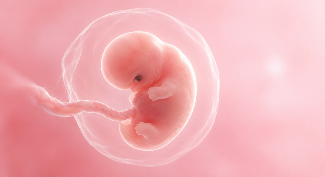
Ever wondered what exactly happens in the earliest days of life? An embryo is the very beginning of human development — a tiny but powerful stage where all major organs, the heart, brain, limbs, and more begin to form. In this article, we explore what an embryo is, how it forms, its development stages week by week, and why this stage matters in fertility and IVF treatment. Whether you are planning a pregnancy or simply curious about human biology, this complete guide will walk you through everything you need to know about embryonic development.
An embryo is the earliest stage of a developing multicellular organism. In humans, it refers to the developing organism from the moment of fertilization through the end of the 8th week of pregnancy. During this critical period, all major body structures and organ systems begin to form. After the 8th week, the developing organism is referred to as a fetus.
1. Definition of an Embryo
The word embryo comes from the Greek word embryon, meaning "that which grows." In biology, an embryo refers to the early developmental stage of a multicellular organism — the phase during which a single fertilized cell grows into a complex, multi-organ structure.
In human biology, an embryo is the developing human from the moment a sperm fertilizes an egg (forming a zygote) until the completion of the 8th week after fertilization. This stage is called the embryonic period and is one of the most critical phases in human development because all the fundamental body systems are established during this time.
2. How Does an Embryo Form?
Human embryo development begins with fertilization — when a single sperm cell penetrates and merges with an egg cell (ovum) in the fallopian tube. This union creates a zygote, a single-celled structure containing the complete genetic blueprint of a new human being — 46 chromosomes in total (23 from the mother, 23 from the father).
Within 24 hours of fertilization, the zygote begins to divide rapidly. It travels down the fallopian tube toward the uterus while continuing to divide. By the time it reaches the uterus — approximately 5 to 6 days later — it has grown into a blastocyst, a hollow ball of around 70 to 100 cells. The blastocyst then implants into the uterine wall (endometrium), and the embryonic stage officially begins.
3. Stages of Embryonic Development
Embryonic development is a carefully organized sequence of events. Here is a week-by-week overview of what happens:
Days 1–3: Zygote and Cleavage
The fertilized egg undergoes rapid mitotic divisions called cleavage. It divides into 2, 4, 8, and then 16 cells, forming what is called a morula — a tiny, solid ball of cells traveling toward the uterus.
Days 4–5: Blastocyst Formation
The morula develops a fluid-filled cavity and transforms into a blastocyst. An inner cell mass forms inside — these cells will become the embryo itself. The outer layer of cells, called the trophoblast, will eventually form the placenta.
Days 6–10: Implantation
The blastocyst burrows into the uterine lining. The hormone hCG is released, signaling pregnancy to the body. This is the hormone detected by home pregnancy tests.
Weeks 2–3: Gastrulation
The embryo reorganizes into three distinct cell layers — the ectoderm, mesoderm, and endoderm. These layers, called germ layers, are the source of every tissue and organ in the human body.
Weeks 3–8: Organogenesis
All major organs begin to form, including the heart, brain, lungs, liver, kidneys, eyes, and limbs. The heart starts beating around Week 4 to 5. By the end of Week 8, the embryo is approximately 1.2 inches long and transitions into the fetal stage.
4. The Three Germ Layers
During gastrulation, the embryo forms three primary germ layers. Each layer gives rise to specific organs and tissues:
Ectoderm (Outer Layer)
The ectoderm forms the nervous system, skin, hair, nails, and sense organs such as the eyes and ears. The brain and spinal cord both originate from the ectoderm.
Mesoderm (Middle Layer)
The mesoderm forms the heart, muscles, bones, kidneys, blood vessels, and the entire circulatory system. It is also responsible for the connective tissues throughout the body.
Endoderm (Inner Layer)
The endoderm forms the lining of the digestive tract, lungs, liver, pancreas, and other internal organs. It gives rise to structures that interact with the body's internal and external environment.
5. Embryo vs. Fetus: Key Differences
The terms embryo and fetus are often used interchangeably, but they refer to two distinct stages of prenatal development. Understanding the difference is important:
Time Period: An embryo exists from fertilization through Week 8. A fetus refers to the developing baby from Week 9 through birth.
Primary Activity: During the embryonic stage, the focus is on organ formation (organogenesis). During the fetal stage, the focus shifts to organ growth and maturation.
Size: An embryo grows from a microscopic single cell to about 1.2 inches. A fetus grows from 1.2 inches to approximately 20 inches at birth.
Appearance: Early embryos do not appear recognizably human. As the fetal stage progresses, features become increasingly human-like.
Sensitivity: The embryonic stage is the most sensitive period for exposure to harmful substances (teratogens) because organs are forming. The fetal stage is still sensitive but slightly less so.
6. Important Embryonic Structures
Several key structures develop alongside the embryo to protect and nourish it:
Amniotic Sac
A fluid-filled sac surrounding the embryo that protects it from physical shocks, maintains stable temperature, and allows movement of the developing limbs.
Placenta
A temporary organ that develops from the outer cells of the blastocyst. It serves as the lifeline between mother and embryo, delivering oxygen and nutrients, removing waste, and producing hormones like progesterone and hCG to maintain the pregnancy.
Umbilical Cord
Connects the embryo to the placenta. It contains two arteries and one vein that carry blood, ensuring a continuous flow of nutrients and oxygen.
Yolk Sac
Present in the earliest weeks, the yolk sac assists in early nutrient transfer and blood cell production before the placenta is fully functional.
7. Significance in Science and Medicine
The embryonic stage is among the most studied periods in biology and medicine. Understanding embryo development has transformed reproductive medicine, genetics, and clinical care.
Embryonic Stem Cell Research
Embryonic stem cells are pluripotent — meaning they can develop into virtually any cell type in the body. This makes them valuable for regenerative medicine and research into diseases such as Parkinson's disease, diabetes, and spinal cord injuries.
Teratology — Study of Birth Defects
Because all major organs form during the embryonic stage, this is when the developing organism is most vulnerable to teratogens — substances or agents that disrupt normal development and can cause birth defects. These include alcohol, certain medications, radiation, and infections like rubella.
Prenatal Diagnosis
Advanced ultrasound and genetic testing can now monitor embryo development and detect chromosomal abnormalities, such as Down syndrome, as early as Week 10 through Non-Invasive Prenatal Testing (NIPT).
8. Embryos in IVF and Assisted Reproduction
In Vitro Fertilization (IVF) has brought embryology directly into clinical practice. In IVF, eggs are retrieved from the ovaries and fertilized by sperm in a laboratory. The resulting embryos are cultured for 3 to 5 days — often to the blastocyst stage — before being transferred into the uterus.
The success rate of a single embryo transfer in women under 35 is approximately 40 to 50 percent per cycle. Embryo quality, assessed through careful grading by embryologists, plays a key role in selecting the most viable embryo for transfer.
Preimplantation Genetic Testing (PGT) is used to screen embryos for chromosomal abnormalities before transfer, improving success rates and reducing the risk of miscarriage. Unused embryos can be frozen (cryopreserved) for future use.
9. Fascinating Facts About Embryos
• The embryonic heart begins beating around 22 days after fertilization — often before a person even knows they are pregnant.
• From the moment of fertilization, the embryo carries a unique DNA sequence — a complete genetic identity different from both parents.
• Early embryos of humans, fish, chickens, and reptiles look remarkably similar, reflecting shared evolutionary ancestry.
• The neural tube — the precursor to the brain and spinal cord — closes by Week 4, which is why adequate folic acid intake before and during early pregnancy is so important.
• All human embryos initially develop with female characteristics. Male sexual differentiation begins around Week 6 to 7, triggered by the SRY gene on the Y chromosome.
Disclaimer
The information provided in this article is intended for general educational and informational purposes only. It does not constitute medical advice, diagnosis, or treatment recommendations.
Every pregnancy and individual health situation is unique. If you have questions or concerns about embryo development, fertility, or pregnancy, please consult a qualified healthcare professional such as a gynecologist, obstetrician, or reproductive specialist.
Do not make decisions about medications, supplements, or fertility treatments based solely on general content. Always seek guidance from a licensed medical professional before starting, stopping, or changing any aspect of your health care.
Medical knowledge evolves continuously. While every effort has been made to ensure the accuracy of this content, readers are encouraged to verify information with up-to-date medical sources and consult their healthcare provider for advice specific to their situation.
